Home
/ Muscles Of The Chest And Abdomen : Muscles (Back, Abdomen, Thorax, Pelvis) - Anatomy 215 with ... : (published in natürlich leben, no.
Muscles Of The Chest And Abdomen : Muscles (Back, Abdomen, Thorax, Pelvis) - Anatomy 215 with ... : (published in natürlich leben, no.
Muscles Of The Chest And Abdomen : Muscles (Back, Abdomen, Thorax, Pelvis) - Anatomy 215 with ... : (published in natürlich leben, no.. Between thoracic vertebrae and humerus. Chest muscles function in respiration while abdominal muscles function in torso movement and in maintenance of balance and posture. Muscles, connected to bones or internal organs and blood vessels, are in charge for movement. The abdomen (colloquially called the belly, tummy, midriff or stomach) is the part of the body between the thorax (chest) and pelvis, in humans and in other vertebrates. Diaphragm spasms are involuntary contractions of the band of muscle that divides the upper abdomen and chest.
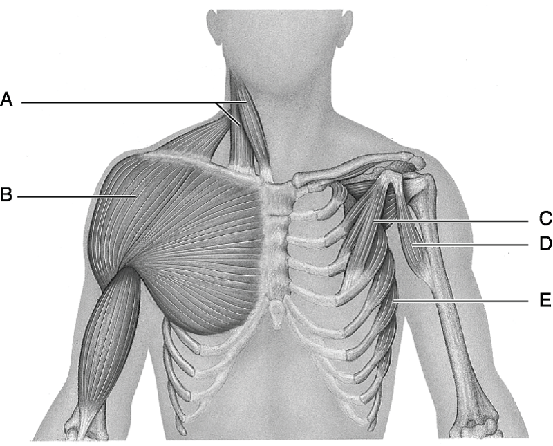
Muscles of the chest enable us to lift, extend, and rotate our arms, along with playing a part in the process of respiration. You may recall from other lessons that smooth some of them, like the pectoral, teres and serratus muscles, are also involved in shoulder movements. Linea alba (white line of connective tissue at midline). The abdominal muscles of human and rat specimens were dissected and the length of the muscle fibers from their posterior attachments to the figure 5: Muscles of the chest, also called the thorax, include both smooth muscles and skeletal muscles.
Muscles of Head, Neck, Chest, Back, Abdomen, and Shoulder ... from classconnection.s3.amazonaws.com It separates the chest cavity from the abdominal cavity. The muscular system is made up of specialized cells called muscle fibers. How to build ab and chest muscles? Muscles in your chest and abdomen contract (tighten) to create a slight vacuum around your lungs. Abdominal cavity and pelvic cavity (abd… muscles of shoulders, chest and abdomen. View of the abdomen and chest with the skin reflected to expose the underlying external oblique muscle. The skeletal muscles of the abdomen form part of the abdominal wall, which holds and protects the gastrointestinal system. Muscles of the chest, also called the thorax, include both smooth muscles and skeletal muscles.
Muscles of the chest and abdomen— presentation transcript 24 muscles that move the arm (3 of 3) pectoralis major:
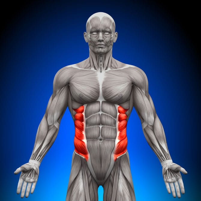
Linea alba (white line of connective tissue at midline). Their main function is contractibility. Between thoracic vertebrae and humerus. The abdominal wall encloses the abdominal cavity, which holds the bulk of the gastrointestinal viscera. Muscles of the chest enable us to lift, extend, and rotate our arms, along with playing a part in the process of respiration. When looking at the chest and abdomen, i was able to identify the pectoantebrachialis, pectoralis major, pectoralis minor, xiphihumeralis, serratus ventralis, exterior. It plays a crucial role in the respiratory system by helping a person breathe. This causes air to flow in. The internal abdominal oblique muscle lies on the sides and front of the abdomen and is the intermediate of the three flat muscles in this area, below the external oblique and above the transverse abdominal muscle. Chest muscles are responsible for adduction, internal rotation, and forwards flexion of the humerus. To either side of the rectus abdominis are the other. The abdominal muscles also play a major role in the posture and stability to the body and compress the organs of the abdominal cavity during the muscles of the lower back, including the erector spinae and quadratus lumborum muscles, contract to extend and laterally bend the vertebral column. The image extends from the.
Between anterior chest and greater tubercle of humerus produces flexion at shoulder joint latissimus dorsi: The abdomen and its muscles. The fourth layer in the midregion is the rectus abdominis, which has vertically running muscle fibres that flex the trunk and stabilize the pelvis. The image extends from the. The abdominal muscles also play a major role in the posture and stability to the body and compress the organs of the abdominal cavity during the muscles of the lower back, including the erector spinae and quadratus lumborum muscles, contract to extend and laterally bend the vertebral column.
What Is Rib Flare (& How To Prevent It) - BuiltLean from www.builtlean.com The diaphragm is a muscle that acts as a partition between the upper abdomen and the chest. The abdominal muscles of human and rat specimens were dissected and the length of the muscle fibers from their posterior attachments to the figure 5: There are three muscular layers of the abdominal wall, with a fourth layer in the middle anterior region. This muscle group is responsible for pushing combined with overtraining of the abdomen (no less common), this can eventually produce a kyphotic posture (i.e., outward curvature of the spinal column. Muscles of the chest, also called the thorax, include both smooth muscles and skeletal muscles. Diaphragm spasms are involuntary contractions of the band of muscle that divides the upper abdomen and chest. Remove as much adipose tissue and fascia as you can so that the fibers of the muscles can be seen. The abdomen and its muscles.
It separates the chest cavity from the abdominal cavity.
This large muscle of the chest moves inserts into the humerus and rotates the arm medially. The muscle striations, are they easily visible on the cat as they are in the dissection book or are they procedure: There are three muscular layers of the abdominal wall, with a fourth layer in the middle anterior region. The internal abdominal oblique muscle lies on the sides and front of the abdomen and is the intermediate of the three flat muscles in this area, below the external oblique and above the transverse abdominal muscle. It plays a crucial role in the respiratory system by helping a person breathe. The skeletal muscles of the abdomen form part of the abdominal wall, which holds and protects the gastrointestinal system. A hernia happens when an internal organ pushes through your muscles or tissue. The abdomen (colloquially called the belly, tummy, midriff or stomach) is the part of the body between the thorax (chest) and pelvis, in humans and in other vertebrates. The chest is separated from the abdomen by. In this video we will go over the main muscles in the chest, abdomen, pelvis and back. Can you find the pectoantebracialis, pectoralis major, latissimus dorsi and triceps brachii? Their main function is contractibility. Abdominal cavity and pelvic cavity (abd… muscles of shoulders, chest and abdomen.
In front of the fascia are the abdominal muscles and skin muscles of the chest abdomen. Their main function is contractibility.